Home
/ Simple Compact Bone Diagram : Schematic Diagram of Compact and Spongy Bones. Schematic ... - A diagram of the anatomy of a bone, showing the compact bone.
Simple Compact Bone Diagram : Schematic Diagram of Compact and Spongy Bones. Schematic ... - A diagram of the anatomy of a bone, showing the compact bone.
Simple Compact Bone Diagram : Schematic Diagram of Compact and Spongy Bones. Schematic ... - A diagram of the anatomy of a bone, showing the compact bone.. However, they do contain osteons, which are like canals, providing passageways through the hard bone matrix. Long bones such as the femur contain two distinct morphological types of bone: Due to its function, compact bone is also referred to as strong bone; B) parallel to the epiphysis. These are shown in the figure below.
Compact bone, also called cortical bone, dense bone in which the bony matrix is solidly filled with organic ground substance and inorganic salts, leaving only tiny spaces (lacunae) that contain the osteocytes, or bone cells.compact bone makes up 80 percent of the human skeleton; Deep to the compact bone layer is a region of spongy bone where the bone tissue grows in thin columns called. The remainder is cancellous bone, which has a spongelike appearance with numerous large spaces and is found in the. B) parallel to the epiphysis. Compact and cancellous — or spongy — bone are the two types of tissue found within most bones.
File:624 Diagram of Compact Bone-new.jpg - Wikimedia Commons from upload.wikimedia.org The fishbone template is a simple visualization of a problem's causes and as the name implies, the diagram has the appearance of a fish skeleton, where each bone represents a category of a root cause. Compact bone forms the outer 'shell' of bone. A diagram of the anatomy of a bone, showing the compact bone. As seen in the image below, compact bone forms the cortex, or hard outer shell of most bones in the body. The remainder of the bone is formed by cancellous or spongy bone. Fracture in which the bone is exposed to the outside Structure of human bones explained. Compact bone, also called cortical bone, dense bone in which the bony matrix is solidly filled with organic ground substance and inorganic salts, leaving only tiny spaces (lacunae) that contain the osteocytes, or bone cells.compact bone makes up 80 percent of the human skeleton;
Compact bone, also called cortical bone, dense bone in which the bony matrix is solidly filled with organic ground substance and inorganic salts, leaving only tiny spaces (lacunae) that contain the osteocytes, or bone cells.compact bone makes up 80 percent of the human skeleton;
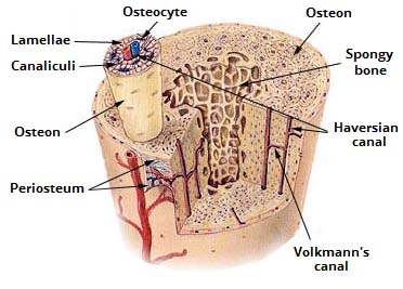
Compact bone forms the outer 'shell' of bone. Related posts of compact bone diagram labeled anatomical diagram of the abdomen. The diagram above shows a longitudinal view of an osteon. A diagram of the anatomy of a bone, showing the compact bone. In this type of bone, the lamellae are organised into concentric circles, which surround a vertical haversian canal (which transmits small neurovascular and lymphatic vessels). Which bone cell in the diagram below is a mature bone cell that helps maintain bone tissue? Compact bone accounts for 80% of the bones in the human body. (b) in this micrograph of the osteon, you can clearly see the concentric lamellae and central canals. The labels include periosteum, compact bone, nutrient artery & vein, medullary cavity, yellow bone marrow, endosteum, epiphyseal line, and spongy bone with red bone marrow. Diagram of a typical long bone: The main type of bone cell is the osteocyte (bone cell, shown as purple in the diagram). Compact bone is made of a matrix of hard mineral salts reinforced with tough collagen fibers. (b) in this micrograph of the osteon, you can clearly see the concentric lamellae and central canals.
Compact bone definition compact bone, also known as cortical bone, is a denser material used to create much of the hard structure of the skeleton. (b) in this micrograph of the osteon, you can clearly see the concentric lamellae and central canals. This provides the bones strength and consists of tightly stacked layers of bone which appear to form a solid section. Due to its structure, it is referred to as cortical bone. Compact bone is the denser, stronger of the two types of osseous tissue (figure 6.3.6).
Supportive connective tissue: Cartilage and Bone - Online ... from www.onlinebiologynotes.com The fishbone template is a simple visualization of a problem's causes and as the name implies, the diagram has the appearance of a fish skeleton, where each bone represents a category of a root cause. In long bones, as you move from the outer cortical compact bone to the inner medullary cavity, the bone transitions to spongy bone. E) it occurs at a faster rate in compact bone than spongy bone. (b) in this micrograph of the osteon, you can clearly see the concentric lamellae and central canals. Anatomical diagram of the abdomen 12 photos of the anatomical diagram of the abdomen anatomical diagram of the abdomen, diagram of the abdomen area, diagram of the abdomen female, diagram of the abdomen muscles, diagram of the abdominal aorta, human anatomy, anatomical diagram of the abdomen, diagram of the. _____ _____ is the hard and dense, but not solid, bone tissue that is beneath the outer membrane of a bone spongy bone ______ _____ is the layer of bone tissue having many small spaces and found just inside the layer of compact bone These are shown in the figure below. Fracture protected by uninjured skin (or mucous membrane) compound (open) fracture:
A diagram of the anatomy of a bone, showing the compact bone.
C) it involves bone deposition. Compact and cancellous — or spongy — bone are the two types of tissue found within most bones. (b) in this micrograph of the osteon, you can clearly see the concentric lamellae and central canals. (b) in this micrograph of the osteon, you can clearly see the concentric lamellae and central canals. However, they do contain osteons, which are like canals, providing passageways through the hard bone matrix. In compact bone, these cells are embedded within the solid calcium phosphate matrix of solid bone. This entire structure is called an osteon and is the functional unit of bone. Learn vocabulary, terms, and more with flashcards, games, and other study tools. It makes up the outer cortex of all bones and is in immediate contact with the periosteum. They are roughly cylindrical, and about 0.2mm wide and a few millimeters long. Due to its function, compact bone is also referred to as strong bone; Osteons are the small units of which the hardest parts of human bones are made. Bone is made up of two base componets:
Which bone cell in the diagram below is an osteogenic cell? D) it occurs at different rates at different locations. Long bones such as the femur contain two distinct morphological types of bone: (b) in this micrograph of the osteon, you can clearly see the concentric lamellae and central canals. The main type of bone cell is the osteocyte (bone cell, shown as purple in the diagram).
AccessJ: How to Donate Bone Marrow from 2.bp.blogspot.com Diagram of a typical long bone: However, they do contain osteons, which are like canals, providing passageways through the hard bone matrix. A diagram of the anatomy of a bone, showing the compact bone. This entire structure is called an osteon and is the functional unit of bone. Compact bone forms the outer 'shell' of bone. Diagram of distinct morphological types of bone. Bones of pelvis pics 12 photos of the bones of pelvis pics , bone. Compact bone definition compact bone, also known as cortical bone, is a denser material used to create much of the hard structure of the skeleton.
This entire structure is called an osteon and is the functional unit of bone.
However, they do contain osteons, which are like canals, providing passageways through the hard bone matrix. Compact and cancellous — or spongy — bone are the two types of tissue found within most bones. Compact bone is the denser, stronger of the two types of osseous tissue (figure 6.3.6). These are shown in the figure below. Osteons are the small units of which the hardest parts of human bones are made. The labels include periosteum, compact bone, nutrient artery & vein, medullary cavity, yellow bone marrow, endosteum, epiphyseal line, and spongy bone with red bone marrow. Under the periosteum is a thin layer of compact bone (often called cortical bone). Shows compact (cortical) and cancellous (spongy) bone. Deep to the compact bone layer is a region of spongy bone where the bone tissue grows in thin columns called. Many tiny cells called osteocytes live in small spaces in the matrix and help to maintain the strength and integrity of the compact bone. Which of the following activities has the greatest effect on bone remodeling and bone deposition? Due to its structure, it is referred to as cortical bone. D) it occurs at different rates at different locations.
C) it involves bone deposition compact bone diagram. Bones of pelvis pics 12 photos of the bones of pelvis pics , bone.

